Histopathology
It is possible to view our range of machines here. Where little is understood, tireless work in the laboratory means a more granular understanding can be developed and eventually, ‘eureka’ moments are born.
At Bright Instruments, we draw great inspiration from the work of those ‘in the field’, and design our cutting edge sectioning equipment with end-users in mind. These slides are what will ultimately be examined under the microscope, and so their meticulous preparation is no mean feat.
The preparation of histology slides is usually a five stage process; the stages are: fixing, processing, embedding, sectioning and last but by no means least, staining.
Sectioning in particular is important as sufficiently thin slices must be prepared in order to enable clear and accurate observation of the minute cell detail, considering electron microscopy or light microscopy both analyse detailed tissue micro structure.
Sectioning typically involves using instruments called micro tomes – which can be combined with freezing units or if frozen sections are required.
and the thickness of cuts required varies depending on analysis techniques and other factors, such as tissue type.
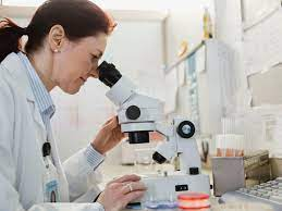
The importance of histopathology cannot be understated – this discipline is absolutely vital to the understanding and detection of diseases, which ultimately broadens and progresses treatment options in the majority of instances. Histopathology is the examination of biological tissues in order to observe the appearance of diseased cells in microscopic detail.
Histopathology typically involves a biopsy, which is a procedure involving taking a small sample of tissue, usually undertaken by a pathologist, who are experts in diagnoses of diseases.
Histopathology involves the preparation of histology slides, which is arguably the most integral element of the process.
Histopathology is study of diseased tissues and cells removed from patients body during an operation. It helps in accurate diagnosis of cancer .
Specimen is processed in a automated tissue processor and sections are placed onto glass slides examined under digital microscope by a pathologist and photographs captured and provided along with the detailed printed report .
Also to ensure the quality performance and to keep random & systemic errors under control , our laboratory has a form of quality management system for all the procedures performed in Histopathology and departments .Quality assurance, continuing quality improvement and quality control are integral components of this system .
To maintain both precision and accuracy performances our laboratory participates in appropriate internal and external quality schemes thereby contributing information for a quality assurance program.
This system is used to approach, evaluate and identify opportunities to improve quality before problems occur through evaluation of all systems/processes in the laboratory.Also specialized in performing special staining on formalin-fixed, paraffin-embedded tissues/cell blocks. These stains are used to detect the presence, abundance, and localization of specific proteins to aid in determining the differentiation of different types of cancers with similar morphology as well as to provide prognostic.
Most commonly performed parameter is staining in cases of breast cancers which plays a vital role in therapy of such cancers .Histopathology is the diagnosis and study of diseases of the tissues, and involves examining tissues and/or cells under a microscope are responsible for making tissue diagnoses and helping clinicians manage a patient's care.
Cytology is often used for preliminary evaluation to establish a working diagnosis and as needed, plan surgery. It is a non-invasive method for gathering preliminary information about certain medical conditions. Little equipment is required and sample collection can often be performed without sedation or anesthesia. The technique can also be used at the time of surgery for on-the-spot assessment of a mass or tumor that has been found.
In some cases, a biopsy it taken knowing that surgical removal of the whole tumor is not possible. In other cases, a small biopsy can be useful to plan the surgical approach or determine if other treatments may be used, thereby increasing the chances of a successful outcome. However, in many cases, particularly when it is easy to get good surgical margins around the mass, it is more appropriate to remove the whole mass. Your veterinarian will give your pet a local or general anesthetic in order to take samples.
The main risks to your pet are either from the disease your pet has or from the anesthetic. In a few cases, particularly when taking biopsies from tumors of the blood vessels, or from organs such as the liver, there can be excessive bleeding.
After the biopsy, your pet should not be allowed to interfere with the biopsy site, which needs to be kept clean and dry. Report any loss of stitches or significant swelling or bleeding to your veterinarian. If you require additional advice on post-surgical care, please ask your veterinary healthcare team. An Elizabethan collar may be recommended to prevent your pet from being able to lick at the incision.
After the biopsy, your veterinarian will place the tissue sample into a preservative solution and submit it to a diagnostic laboratory. In the laboratory, the process of histopathology begins.


Comments
Post a Comment